
10 September 2026 · TJ Smile Studio
Avoid Rework: 30–60 Minute Digital Bite Analysis for Patients
How digital bite analysis with T-Scan finds hidden bite issues, helps prevent restoration rework, and guides precise, evidence-based adjustments.
Digital bite analysis uses a pressure-sensitive sensor and software to record, in milliseconds, exactly how and when your teeth meet as you bite down, plus the relative force at each contact point. That timing and force map gives dentists measurable data, not a visual guess, to diagnose occlusal imbalance and plan adjustments that actually target the problem. The process, its metrics, and the evidence behind it are worth understanding before your next appointment.
TL;DR:
- Digital bite analysis provides precise timing and force data, with an ideal occlusion time under 0.2 seconds and disocclusion time under 0.5 seconds for most cases.
- The assessment is most beneficial for diagnosing jaw pain, bruxism, and ensuring restorations do not absorb excessive force during initial contact.
- Combining digital analysis with traditional methods like articulating paper and intraoral scans yields the most complete understanding of occlusal issues.
- Digital tools produce comparable, trackable results, but interpretation requires experienced clinicians due to device variability and trend analysis complexity.
- Most patients underestimate how small timing and force discrepancies can cause long-term symptoms, emphasizing the need for precise digital diagnostics in treatment planning.
Table of Contents
- What is digital bite analysis and how does it differ from a bite check?
- What happens during a digital bite assessment?
- Which conditions benefit most from digital bite analysis?
- How does it compare with articulating paper, and intraoral scanning?
- What does the research actually show about outcomes?
- How TJ Smile Studio uses digital bite analysis in practice
- What patients get wrong about bite analysis
- Booking a digital bite assessment at TJ Smile Studio
- Sources
- FAQ
What is digital bite analysis and how does it differ from a bite check?
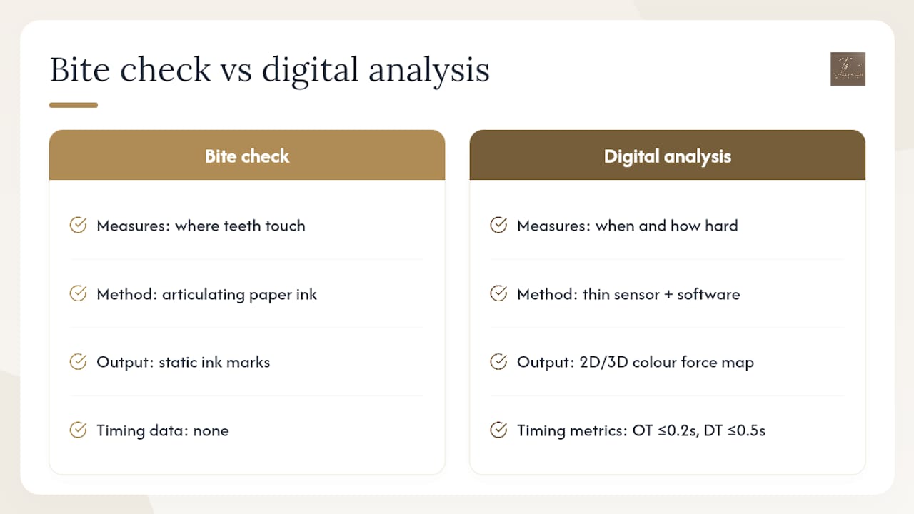
A standard bite check usually means biting on articulating paper and watching where ink marks appear. It tells a dentist where your teeth touch, but nothing about when they touch or how hard. Digital bite analysis, most commonly performed with systems like T-Scan, closes that gap by recording contact sequence and relative pressure in real time on a thin, flexible sensor you bite into.
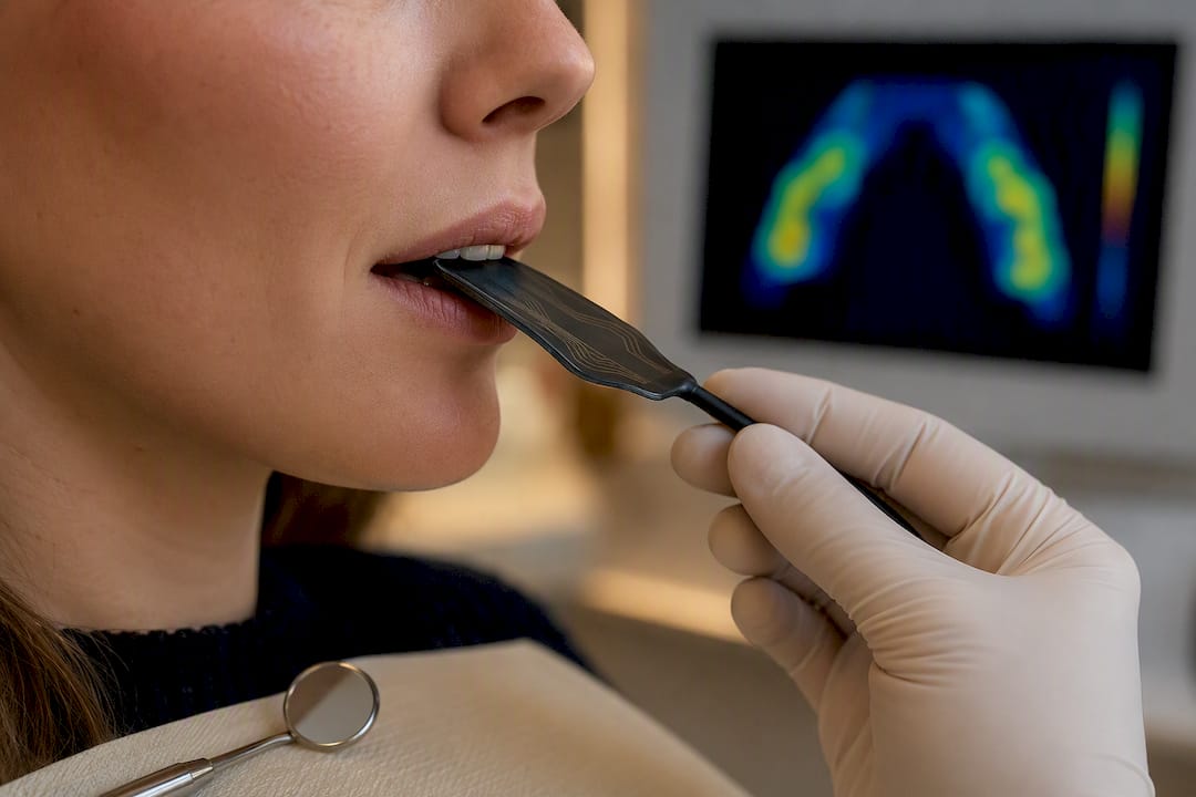
The sensor feeds data to software that displays a 2D or 3D colour map, red for heavy contact, blue or green for lighter contact, updating as you close your jaw. Two numbers matter most here. Occlusion time (OT), the time it takes all your teeth to make contact, ideally sits at or below 0.2 seconds. Disocclusion time (DT), how quickly your back teeth separate during a lateral chewing motion, ideally stays under 0.5 seconds. Longer times on either measure usually point to interfering contacts or muscle strain, and reducing DT has been linked to lower masseter and temporalis muscle hyperactivity in some studies.
Pro Tip: The percentages and colours on a T-Scan display show relative force distribution across your arch, not force in newtons. Think of it as a comparison map, not a scale.
The software also tracks the centre of force (COF), a single point showing where your overall bite force lands, and lets a clinician play back the entire closing motion frame by frame to spot exactly when an imbalance appears.
What happens during a digital bite assessment?
A digital bite assessment usually slots into a broader restorative or occlusal consultation rather than standing alone, and most patients move through a fairly consistent sequence.
- Initial consultation. Your dentist reviews symptoms, jaw history, and any grinding or clicking, then explains why a bite recording is useful for your case.
- Sensor recording. You bite into a thin sensor (comparable in feel to a slim mouthguard) several times while the software captures timing and force data live on screen.
- Complementary records. Many clinicians pair the digital reading with an intraoral scan, using a system such as the Trios scanner from 3Shape, and sometimes a quick articulating paper check to cross-reference static contact points.
- Interpretation. The dentist reviews OT, DT, and the force map together, looking for early contacts, asymmetric loading, or delayed disocclusion.
- Targeted adjustment. Using that data, adjustments are made in small, measured steps, often re-checked with another digital recording immediately after.
The recording itself takes only a few minutes and causes no discomfort since the sensor is thin enough to bite naturally. A full assessment with adjustment or splint fitting typically runs 30 to 60 minutes, with follow-up reviews scheduled over subsequent weeks to confirm the bite has stabilised.
Which conditions benefit most from digital bite analysis?
Digital bite analysis earns its place most clearly in cases where a small, hidden discrepancy in timing or force is driving a much bigger symptom.
- TMJ dysfunction and jaw pain. Uneven disocclusion timing correlates with muscle overactivity, so measuring DT gives a concrete target for treatment rather than relying on patient-reported pain alone.
- Bruxism and occlusal splints. Splints designed and adjusted using digital feedback can be fine-tuned to specific OT/DT values instead of a generalised fit, with documented reductions in both metrics after guided adjustment.
- Implant and crown protection. New restorations that sit even slightly high can absorb excessive force during the first moments of closure. A force map flags that early contact before it causes discomfort or mechanical failure.
- Veneer and full mouth rehabilitation cases. When multiple teeth are restored at once, confirming balanced contact across the whole arch matters more than checking each tooth individually.
- Orthodontic and prosthetic finishing. Digital verification confirms that a finished case actually disoccludes evenly, rather than assuming it based on how it looks.
In each case, the underlying logic is the same: symptoms often trace back to fractions of a second and small percentage differences in force, both of which are invisible without the readout.
How does it compare with articulating paper, and intraoral scanning?
None of these tools replaces the others outright, and the research backs that up rather than favouring one method across the board.
What digital analysis adds:
- Contact timing in milliseconds, something ink marks simply cannot record
- Relative force distribution across the whole arch in one view
- A saved, comparable record for tracking progress over multiple visits
- Visual, playback-based feedback that makes the problem easier to explain to a patient
Where it falls short:
- A genuine learning curve; interpreting force maps and OT/DT trends takes practice
- Higher cost and equipment investment than a pack of articulating paper
- Sensor thickness, thin as it is, can still slightly alter natural bite in some patients
- Readings are comparative, not an absolute force measurement in newtons
Comparative research backs up that trade-off directly: articulating paper often detects more static contact points in certain areas than digital sensors do, even though it captures nothing about timing or force. A separate in vitro comparison across digital analysers and articulating paper found real variability between devices, concluding that no single method matches paper’s static accuracy while also capturing dynamic occlusion well. Inter-rater reliability studies report Cohen’s Kappa values near or above 0.9 for articulating paper, a reminder that the old method still has real strengths.
The practical takeaway from that evidence is straightforward: combine methods. Static paper checks catch contact points, digital analysis catches timing and force, and intraoral scanning ties both to a 3D model for planning.
What does the research actually show about outcomes?
The most concrete evidence comes from a documented case using T-Scan Novus in occlusal splint treatment for bruxism, where a patient’s occlusion time and disocclusion time were both recorded before and after digitally guided adjustment.
The patient’s occlusion time fell from a higher initial value to a significantly lower value after adjustment, approaching clinically desirable targets. Disocclusion time also decreased to more normal levels, which is consistent with reductions in muscle hyperactivity reported in similar cases.
That is a single case report, not a large trial, so it demonstrates a plausible mechanism and a measurable result rather than proof the same improvement happens for everyone. Broader comparative work on inter-rater reliability shows that interpretation still depends heavily on clinician experience, and device-to-device variability means results from one system do not automatically generalise to another. The consistent thread across the research is that digital timing and force data give clinicians something concrete to track, adjust against, and re-measure, which articulating paper alone cannot provide.
How TJ Smile Studio uses digital bite analysis in practice
At TJ Smile Studio in Glasgow, digital bite analysis sits inside a broader digital workflow led by Dr Tashfeen Jamil (BDS, MFDS RCS Ed, AssocFCGDent, GDC 286427). Intraoral scanning captures the full arch digitally, and bite timing and force data are reviewed alongside that model before any restorative or cosmetic work begins, whether that is a same-day smile transformation or a staged full mouth plan.
Patients typically notice three things during this process: a clear baseline reading of their own bite before treatment starts, live visual feedback on screen as adjustments happen, and a follow-up recording that confirms the change actually held. For treatments involving multiple new restorations, such as veneers or bonding across several teeth, that verification step matters, because a bite that looks balanced can still carry an uneven load that only shows up on a force map.
Combining scanning with bite data lets adjustments happen in smaller, more accurate increments rather than repeated trial-and-error visits, which fits naturally with a practice-level digital equipment and workflows approach.
What patients get wrong about bite analysis
Most patients assume a “good bite” means their teeth simply look even and meet without obvious pain. The evidence here says otherwise: timing and force imbalances can sit well below the threshold of conscious discomfort for years before they surface as jaw pain, worn enamel, or a fractured restoration. That is the biggest blind spot in how this topic gets discussed generally, and it is worth prioritising over cosmetic appearance alone when planning restorative work.
The second misconception is treating digital analysis as a replacement for clinical judgement. A force map or OT/DT reading is data, not a diagnosis. It still takes an experienced eye to decide whether a reading reflects a genuine problem or normal individual variation, which is exactly why the reliability research keeps pointing back to clinician training rather than the device alone.
Where the conventional advice falls short is in treating articulating paper and digital analysis as competing choices. They measure different things entirely, and the research is fairly consistent that combining them gives a more complete picture than either alone. If there is one practical priority for a patient, it is this: ask whether timing and force were checked, not just contact points, before agreeing to any bite adjustment or new restoration.
— Dr Tashfeen Jamil BDS, MFDS RCS Ed, AssocFCGDent - GDC 286427
Booking a digital bite assessment at TJ Smile Studio
TJ Smile Studio offers diagnostic digital bite assessment as part of its wider digital smile makeover service in Glasgow, using intraoral scanning alongside bite timing and force data to plan treatment with fewer follow-up adjustments than a purely visual approach. That matters most for anyone considering veneers, bonding, or a fuller restorative plan, where an uneven bite discovered after the fact means redoing work rather than getting it right the first time.
At your first appointment, expect a short consultation covering your dental history and any jaw discomfort, followed by the scan and bite recording itself, both quick and non-invasive. There is no special preparation needed beyond arriving with your natural bite, avoiding chewing gum or anything unusually hard beforehand. From there, Dr Jamil reviews the results with you directly and explains what they mean for your specific treatment options, from composite bonding to a full veneer plan. If you want to see how this fits your case, get in touch to arrange a consultation and diagnostic assessment.
This article is general information, not a substitute for advice from a qualified doctor. Consult a qualified healthcare professional about your own circumstances before acting on anything here.
Sources
- Digital Occlusal Analysis | T-Scan (Tekscan)
- In vitro comparison of digital occlusal analyzers and articulating paper in static and dynamic occlusion (Journal of Dentistry, 2025)
FAQ
What is a digital bite?
A digital bite is a computerised recording of how your teeth meet, capturing contact timing in milliseconds and relative force distribution across your arch, rather than just showing where contact occurred.
What is the 3-3-3 rule for teeth?
This term does not correspond to a recognised clinical standard in occlusal or restorative dentistry; if you have seen it referenced elsewhere, ask your dentist directly what specific guideline they mean.
How much does a digital dental impression cost?
Pricing for digital scanning and bite analysis varies by clinic and by whether it is bundled into a wider treatment plan such as a digital smile makeover, so it is best confirmed at consultation rather than assumed from a generic figure.
What is OPG and IOPA?
OPG (orthopantomogram) is a single wide X-ray showing your full upper and lower jaw, while IOPA (intraoral periapical) is a close-up X-ray of one or two teeth and their roots; both are separate imaging tools from bite analysis but are often taken alongside it for a complete diagnostic picture.
Recommended
This article is general information, not personal dental advice. What’s right for your teeth depends on a clinical examination — the condition of your enamel, gums, bite and any existing dental work — and no article can assess that. Outcomes vary between patients, and all treatment at TJ Smile Studio is subject to assessment and clinical suitability.
- Dr Tashfeen Jamil BDS, MFDS RCS Ed, AssocFCGDent, PGDip Restorative and Aesthetic Dentistry — GDC registration 286427
This article is general information, not personal dental advice. What's right for your teeth depends on a clinical examination — the condition of your enamel, gums, bite and any existing dental work — and no article can assess that. Outcomes vary between patients, and all treatment at TJ Smile Studio is subject to assessment and clinical suitability.
Clinically reviewed by Dr Tashfeen Jamil BDS, MFDS RCS Ed, AssocFCGDent, PGDip Restorative and Aesthetic Dentistry — GDC registration 286427, Principal Dentist at TJ Smile Studio, 1 Spiersbridge Way, Thornliebank, Glasgow G46 8NG.
If you have dental pain, swelling or an injury, call the practice on 07728 808510. Out of hours in Scotland, call NHS 24 on 111.